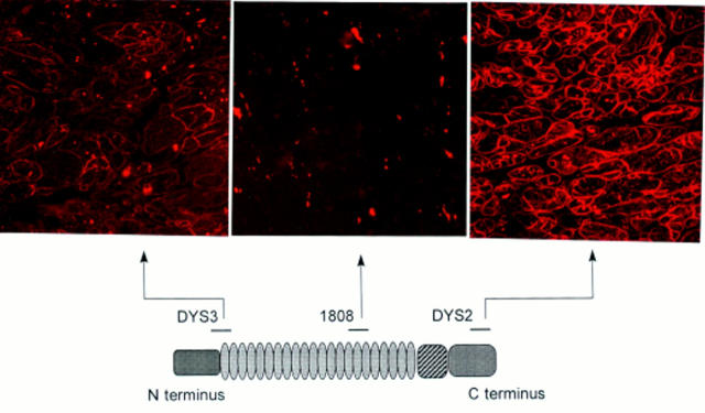
Figure 3 .
Top, immunohistochemical staining of cardiac muscle from patient 2 using antidystrophin antibodies. Bottom, protein dystrophin and epitopes of three of the antibodies used in the study. Top left, N terminus antidystrophin antibodies (Dys3) with a near normal reaction; top middle, no staining was visible with mid-rod domain 1808 antidystrophin antibodies; top right, reduced staining with Dys2 C terminus antidystrophin antibodies (original magnifications ×320).

