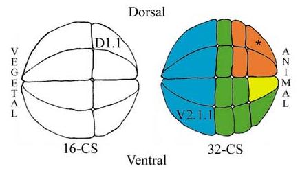
Figure 1.

Side views of 16- and 32-cell stage (CS) Xenopus embryos noting the positions of the two blastomeres manipulated in the described experiments (D1.1, V2.1.1). The 32-cell embryo shows four of the eight blastomeres that are highly retinogenic (orange), and the one that is slightly retinogenic (yellow); asterisk indicates the most retinogenic cell. Several other blastomeres are competent to form retina but normally do not (green) and vegetal blastomeres are not competent to make retina (blue).
