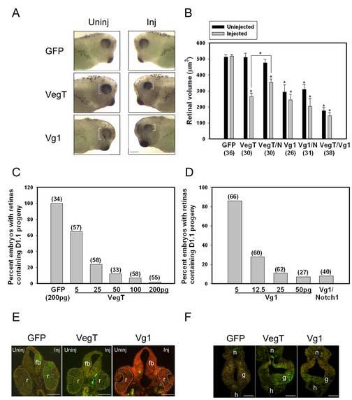
Figure 2.

VegT and Vg1 inhibit the retinal fate of the dorsal-animal (D1.1) blastomere.
(A) Expression of VegT or Vg1 in the D1.1 lineage results in a smaller eye (white brackets). Uninj, uninjected side; Inj, injected side. Scale bar = 400μm.
(B) The mean volume of retinas in embryos in which the indicated mRNAs (GFP, VegT, Vg1, notch1 [N]) were expressed. Numbers in parentheses indicate size of sample. * indicates p<0.01 compared to GFP controls. VegT and Vg1 both significantly reduced the size of the retina on the injected side; Vg1 additionally reduced the retina on the uninjected side. Co-injection of notch1 significantly increased the retinal volume of VegT-injected embryos, compared to VegT alone (bracket, p<0.01), although they remained smaller than the GFp controls. Co-injection of notch1 did not increase the retinal size of Vg1-injected embryos compared to Vg1 alone. Co-injection of VegT and Vg1 mRNAs further reduced the size of the retina compared to VegT alone (p<0.01) or Vg1 alone (p<0.05).
(C) Percentage of embryos in which D1.1 progeny populate the retina. The number is dramatically reduced in a dose-dependent manner in embryos injected with VegT mRNA.
(D) Percentage of embryos in which D1.1 progeny populate the retina. The number is dramatically reduced in a dose-dependent manner in embryos injected with Vg1 mRNA; this is not reversed by co-injection of notch1 (500pg).
(E) The retina (r) on the injected (right) side of VegT and Vg1 embryos is smaller, and the D1.1 progeny (green) that normally contribute large numbers to the retina and forebrain (fb) in GFP controls are missing in VegT and Vg1 embryos. Scale bar = 200μm.
(F) The size of the D1.1 clone (green) in the gut (g) is larger in VegT- and Vg1-injected embryos compared to GFP controls. h, heart; n, notochord. Scale bar = 200μm
