Figure 10.
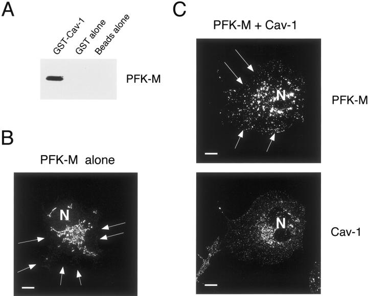
Cav-1 interacts with PFK-M and induces the membrane recruitment of PFK-M. A: GST pull-down assay. 293T cells were transiently transfected with the cDNA encoding PFK-M carrying the V5 epitope tag at its C-terminus. Cell lysates were then incubated with affinity-purified GST alone or GST-Cav-1 (residues 1 to 178) immobilized on glutathione-agarose beads. After extensive washing, the beads were resuspended in 3× SDS-PAGE sample buffer, boiled, and separated by SDS-PAGE. The nitrocellulose membranes were subjected to Western blot analysis with the anti-V5 monoclonal antibody. Note that only GST-Cav-1 binds PFK-M; no binding was observed with GST alone or with the same incubation performed with beads alone. B–C: Immunofluorescence. Cos-7 cells were transfected with the cDNA encoding V5-tagged PFK-M, alone or in combination with Cav-1. Thirty-six hours after transfection, cells were formaldehyde-fixed and immunostained with a monoclonal antibody directed against the V5 epitope (B), or doubly immunostained with the anti-V5 mAb and the anti-Cav-1 pAb (C). Note that when expressed alone, PFK-M is found mostly intracellularly in a perinuclear distribution. Interestingly, recombinant expression of Cav-1 induces the membrane recruitment of PFK-M. N, Nucleus. Scale bars, 10 μm.

