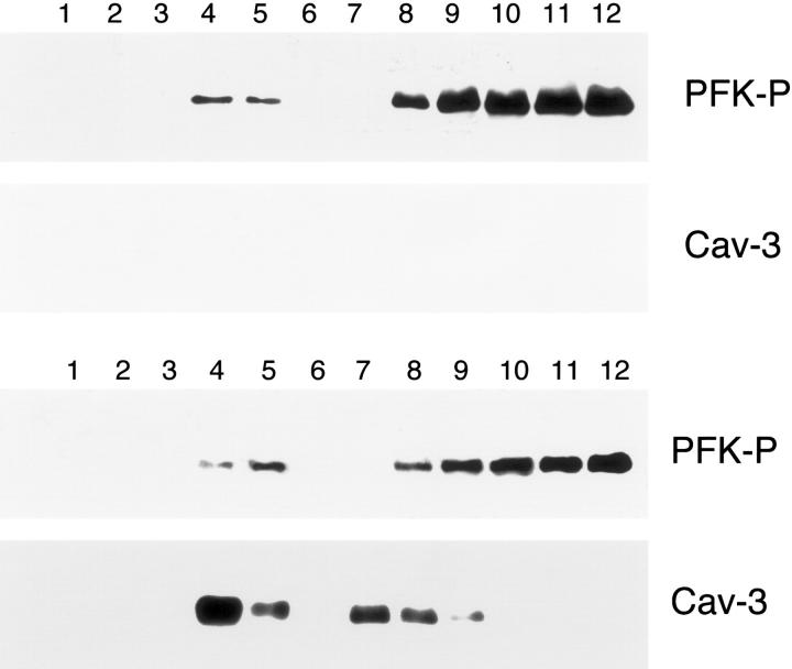
Figure 13.
The expression of Cav-3 does not affect the targeting of PFK-P to lipid rafts/caveolae-enriched fractions. Lipid rafts/caveolae-enriched membranes were separated from other cellular components using an established sucrose density gradient system. In this fractionation scheme, immunoblotting with the anti-Cav-3 mAb probe can be used to track the position of lipid rafts/caveolae-originated membranes (fractions 4 and 5). Cos-7 cells were transiently transfected with the cDNA encoding V5-tagged PFK-P, alone or in combination with Cav-3. Thirty-six hours after transfection, cells were lysed in a buffer containing 1% Triton X-100 and subjected to sucrose gradient centrifugation. Twelve fractions were collected and equal amounts of protein from each fraction were separated by SDS-PAGE and blotted onto nitrocellulose. The distributions of PFK-P and Cav-3 were analyzed by immunoblotting with V5 and Cav-3 antibodies, respectively. Note that the targeting of PFK-P to lipid rafts/caveolae-enriched fractions is not affected by the presence of Cav-3. Interestingly, PFK-P is mostly excluded from lipid rafts/caveolae-enriched domains (fractions 4 and 5) and instead is concentrated at the bottom of the gradient (fractions 8 to 12).

