
Figure 8.
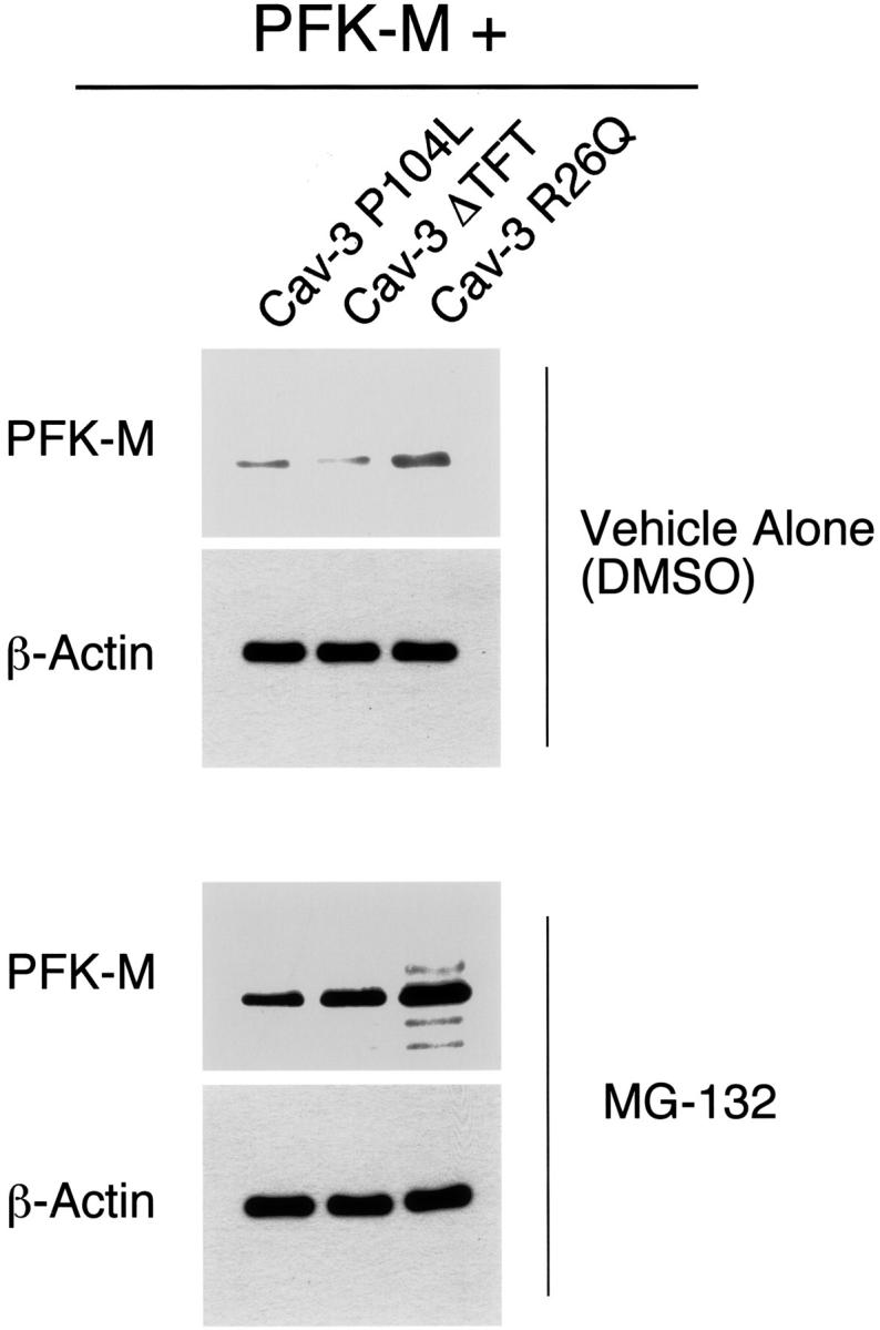
Treatment with the proteasome inhibitor MG-132 rescues the degradation of PFK-M induced by Cav-3 mutants. Cos-7 cells were transiently co-transfected with the cDNAs encoding V5-tagged PFK-M and pathogenic Cav-3 mutants (Cav-3 P104L, Cav-3 ΔTFT, and Cav-3 R26Q). Twenty hours after transfection, cells were incubated with the proteasomal inhibitor MG-132 (10 μmol/L) or with vehicle alone (DMSO). After 16 hours of treatment, the cells were lysed in hot sample buffer and proteins were resolved by SDS-PAGE. Transferred proteins were analyzed by V5 immunoblotting. Note that upon treatment with MG-132, PFK-M expression levels are greatly increased (bottom). In contrast, no effect was observed when cells were treated with vehicle alone (DMSO; top). Note that the V5 blots (top and bottom) were acquired with the same exposure time. Western blot analysis with anti-β-actin IgG is shown as a control for equal loading.