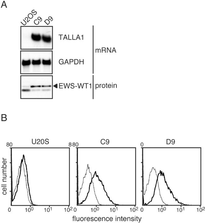
Figure 1.
Expression of Talla-1 in cell clones stably expressing EWS-WT1(-KTS). A: Northern blotting analysis of Talla-1 mRNA. Total RNA (5 μg) was loaded in each lane. EWS-WT1(-KTS) protein (filled triangle) was detected by Western blotting with anti-WT1 antibody. B: Flow cytometric analysis of TALLA-1 protein. Living cells were incubated with anti-TALLA-1 (B2D) monoclonal antibody and Alexa-Fluor 488-labeled anti-mouse IgG and then subjected to flow cytometric analysis (solid line). Background profiles (dotted line) were measured by staining with Alexa-Fluor 488-labeled anti-mouse IgG only.

