Figure 3.
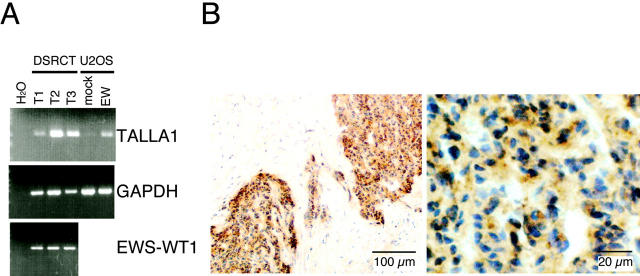
Expression of TALLA-1 in DSRCT specimens. A: RT-PCR analysis of the Talla-1 mRNA in DSRCT. The Talla-1 mRNA was amplified from various sources by RT-PCR and then separated by electrophoresis on agarose gels. Sources are DSRCT tissues (T1, T2, T3), cells transiently expressing EWS-WT1(-KTS) (EW) or its negative control (mock), and various human tissues. Expression of EWS-WT1 fusion transcript was also detected in the three DSRCT tumors. PCR products were visualized by staining with ethidium bromide and UV transillumination. B: Immunohistochemical staining of a DSRCT specimen (T3). TALLA-1-positive cells are stained brown. Sections were counter-stained with hematoxylin.

