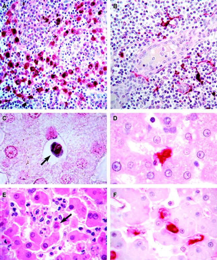
Figure 10.

Localization of EBOV in cynomolgus monkey tissues. A: Immunopositive mononuclear cells (red) in the medullary sinus of a lymph node at day 3. The brown-pigmented cells are hemosiderin-laden macrophages. B: Immunopositive (red) dendritic cells surrounding a high endothelial venule in a lymph node at day 4. C: EBOV RNA-positive circulating monocyte (arrow) in hepatic sinusoid at day 2. D: Immunopositive (red) Kupffer cell at day 3. E: Histology of liver showing small foci of hepatocellular degeneration and necrosis and foci of pleomorphic eosinophilic intracytoplasmic inclusions (arrows) in hepatocytes at day 5. F: Immunopositive Kupffer cells (red) and hepatocytes (red) at day 5. Alkaline phosphatase method, A, B, D, F; H&E stain, E. Original magnifications: ×20 (A); ×40 (B); ×60 (C to F).
