
Figure 11.
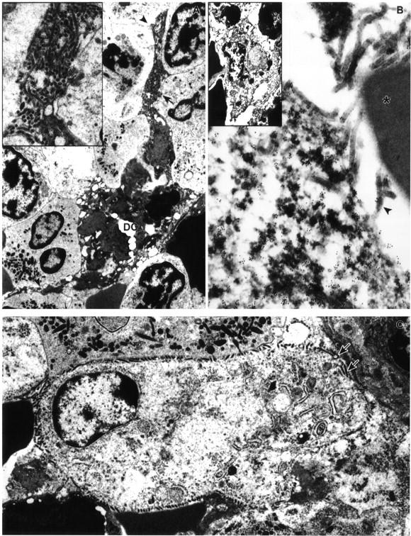
Ultrastructural appearance of dendritic cells in marginal zone of spleen of EBOV-Zaire-infected cynomolgus monkeys. A: EBOV-infected dendritic cell at day 4 with typical branching processes (arrowheads). Inset: Enlargement of area marked by arrow in A shows virions budding from plasma membrane. B: Immunoelectron microscopy showing positive gold sphere (10 nm) labeling of plasma membrane and cytoplasm of EBOV-infected dendritic cell for DC-SIGN at day 3. Note virions budding from plasma membrane (arrowhead) and near absence of gold spheres on adjacent red blood cell (*). Inset: Low-power view of EBOV-infected, DC-SIGN-positive pale-staining cell interpreted as an immature dendritic cell. C: EBOV-infected pale-staining cell interpreted as an immature dendritic cell at day 3. Morphology is comparable to DC-SIGN positive cell in inset of B. Note virions budding from plasma membrane (arrows), pale-staining cytoplasm, and sparsity of organelles. Original magnifications: ×6,500 (A and B, inset); ×12,500 (C); ×16,000 (A, inset); ×53,000 (B).