
Figure 12.
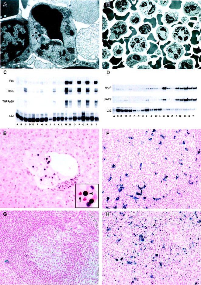
Analysis of PBMC from EBOV-Zaire-infected cynomolgus monkeys for evidence of apoptosis. A and B: Transmission electron micrographs of peripheral blood mononuclear cells. A: Apoptosis of a large lymphocyte (∼6 × 8 μm in cross-section) morphologically consistent with a NK cell at day 3. Arrowheads indicate cytoplasmic granules. Also, note abundance of mitochondria. B: Apoptotic small lymphocyte (arrow) and increased numbers of neutrophils at day 4. C and D: Analysis of PBMC mRNA; representative RNase protection assays are shown. C: Fas and TRAIL, Comparison of preinfection PBMC (lanes A, D, H, K, O, Q, S) with PBMC at postinfection day 1 (lanes B and E); day 2 (lanes F, I, L); day 3 (lanes C, G, J); day 4 (lane M); day 5 (lanes P and R); and day 6 (lanes N and T). D: NAIP and cIAP2. Comparison of preinfection PBMC (lanes A to C) with PBMC at postinfection day 1 (lanes D to F); day 2 (lanes G to I); day 3 (lanes J to L); day 4 (lanes M to O); day 5 (lanes P to R); and day 6 (lanes S and T). E to H: Analyses of tissues by TUNEL assay. E: Apoptotic mononuclear cells (black reaction product) within the lumen of a central hepatic vein and to the periphery of the vein at day 3. Arrow in the inset points to an unaffected neutrophil and the arrowhead indicates the apoptotic lymphocyte. At day 4, follicles in a lymph node contain macrophages and dendritiform cells that have engulfed tingible bodies (F). G: A follicular area in spleen of an uninfected control monkey, showing little evidence of apoptosis, compared to a similar area in spleen of an EBOV-infected monkey at day 5, showing macrophages that have engulfed tingle bodies; single-strand breaks appear blue/black in F to H. Original magnifications: ×20 (E to H); ×6,500 (B); ×16,000 (A).