
Figure 5.
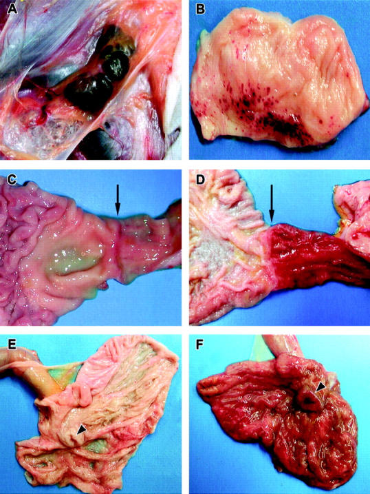
Representative gross necropsy lesions from cynomolgus monkeys experimentally infected with EBOV-Zaire. A: Mild enlargement and marked congestion/hemorrhage of inguinal lymph nodes at day 4. B: Multifocal to coalescing hemorrhages of mucosa of urinary bladder at day 5. C and D: Progression of marked congestion of the duodenum occurring between day 3 (C) and day 5 (D). Arrows indicate the gastroduodenal junction demarcating the stomach to the left and the duodenum to the right. The duodenum is markedly congested at day 5 (D). E and F: Progression of congestion of cecum occurring between day 3 (E) and day 5 (F). The cecum is opened up and the ileum extends outward from the cecum. Arrowheads indicate the ileocecal junction. Note the congested and thickened appearance of the cecum at day 5 (F).