
Figure 2.
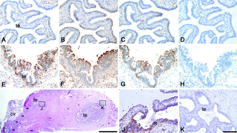
Expression of trophinin, tastin, and bystin proteins in intact fallopian tube and in fallopian tube with tubal pregnancy. Immunohistochemistry of an intact fallopian tube (A–D) and a fallopian tube with tubal pregnancy (E–K) for trophinin (A, E, J, K), tastin (B, F), and bystin (C, G). Hematoxylin and eosin-stained specimen from a tubal pregnancy (I), and high magnification of insets showing immunohistochemistry for trophinin in the area close to (J) or distant from (K) the chorionic villi. The implantation site in (I) is marked by arrowheads. All photographs except (I) are in the same magnification, and the bar in (K) indicates 100 μm. Bar in (I) indicates 2.0 mm. (cv, chorionic villi; te, tubal epithelia).