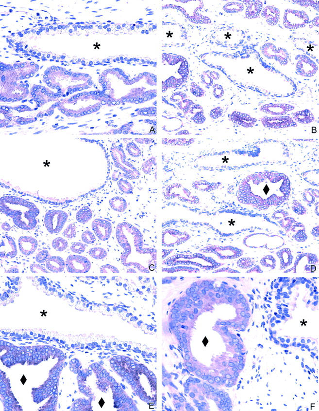
Figure 1.

EphA2 immunohistochemistry in the prostate. A to D: Normal prostate glands show no or minimal staining in the secretory cells lining the lumen of the gland. Adjacent cancer cells show strong expression. E to F: EphA2 expression in high-grade prostatic intraepithelial neoplasia (PIN). The adjacent normal gland showed no or minimal EphA2 immunoreactivity. ♦, PIN; *, adjacent normal glands.
