Figure 2.
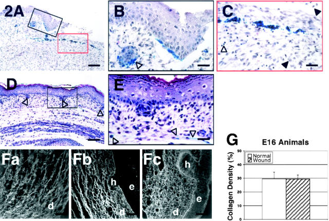
H&E stain of wounded E16 rat skin. A: 24 hours post-injury; magnification, ×100. There is minimal inflammatory infiltrate. B: 24 hours post-injury; magnification, ×400. The presence of blue vital dye in hair follicles near the migrating epithelial edge suggests concurrent hair follicle regeneration with wound re-epithelialization (black open arrow). C: 24 hours post-injury; magnification, ×400. Neutrophils (black solid arrows) and lymphocytes (black open arrow) are the predominant cells of the wound periphery and the center of the wound, respectively. D: 72 hours post-injury; magnification, ×100. The wound is entirely healed with complete regeneration of the normal skin architecture. Normal distribution of hair follicles (black open arrows) are observed in the dermis. E: 72 hours post-injury; magnification, ×400. At higher magnification, the previous wound site, as indicated by the presence of blue vital stain in the dermis (black open arrows) is indistinguishable from the non-wounded skin. F: Confocal microscopic view of wounded E16 rat skin and control. (Fa through Fc). Fa: 48 hours post-injury; magnification, ×630. Note the organized appearance of the collagen fibers with a reticular lattice structure. Fb: 72 hours post-injury; magnification, ×630. The wound site is completely re-epithelialized with complete restoration of normal skin collagen architecture and hair follicle regeneration. Fc: Non-wounded E19 skin [ie, E16 + 72 hours]; magnification, ×630. No difference is observed between E16 skin, 72 hours post-wounding, and non-wounded E19 skin. e, epidermis; h, hair follicle; d, dermis. Bars: A and D, 200 μm; B, C, and E, 50 μm; Fa-Fc, 32 μm. G: There is no significant difference in total collagen density between E16 fetuses 72 hours post-injury and non-wounded E19 (E16 + 72 hours) fetuses (P >0.05).

