
Figure 3.
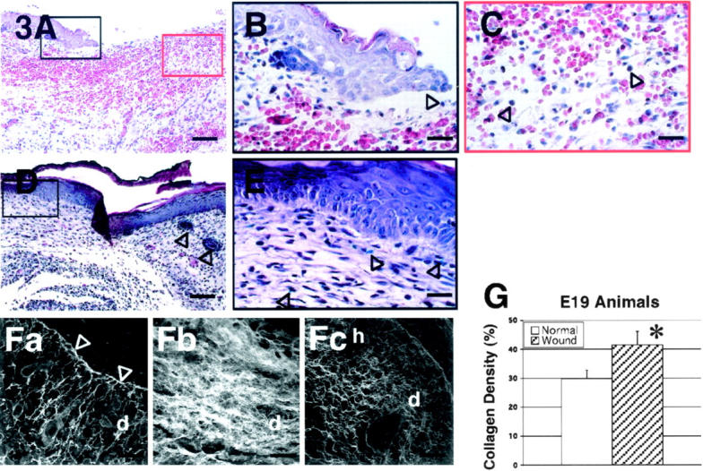
H&E stain of wounded E19 rat skin. A: 24 hours post-injury; magnification, ×100. There is moderate inflammatory infiltrate and increased red blood cells. B: 24 hours post-injury; magnification, ×400. Re-epithelialization is also noted at 24 hours after injury (black open arrow). C: 24 hours post-injury; magnification, ×400. Monocytes (black open arrows) comprises most of the inflammatory cells. D: 72 hours post-injury; magnification, ×100. The wound is completely re-epithelialized with increased cellularity and neovascularity. Hair follicles (black open arrows) are not observed in the repaired wound site (far left) compared with unwounded site (far right). E: 72 hours post-injury; magnification, ×400. At higher magnification, blue vital dye (black open arrows) within the repaired wound is visible. F: Confocal microscopic view of wounded E19 rat skin and control. (Fa through Fc). Fa: 48 hours post-injury; magnification, ×630. Large spaces among newly formed collagen fibers within the dermis are noticeable. A thin layer of dense collagen fibers is seen as basement membrane (white open arrows). Fb: 72 hours post-injury, magnification, ×630. Disorganized collagen deposition pattern with heterogeneously sized collagen fibers is apparent in the healed dermal scar tissue. Note the absence of hair follicle regeneration. Fc: Non-wounded neonatal day 1 (N1) skin [ie, E19 + 72 hours, E21 = term]; magnification, ×630. Non-wounded N1 skin exhibited an organized collagen deposition pattern that is significantly different from E19 skin, 72 hours post-wounding. e, epidermis; h, hair follicle; d, dermis. Bars: A and D, 200 μm; B, C, and E, 50 μm; Fa-Fc, 32 μm. G: There is significantly increased total collagen density in wounded E19 fetuses 72 hours post-injury relative to non-wounded N1 controls (P = 0.00043).