
Figure 4.
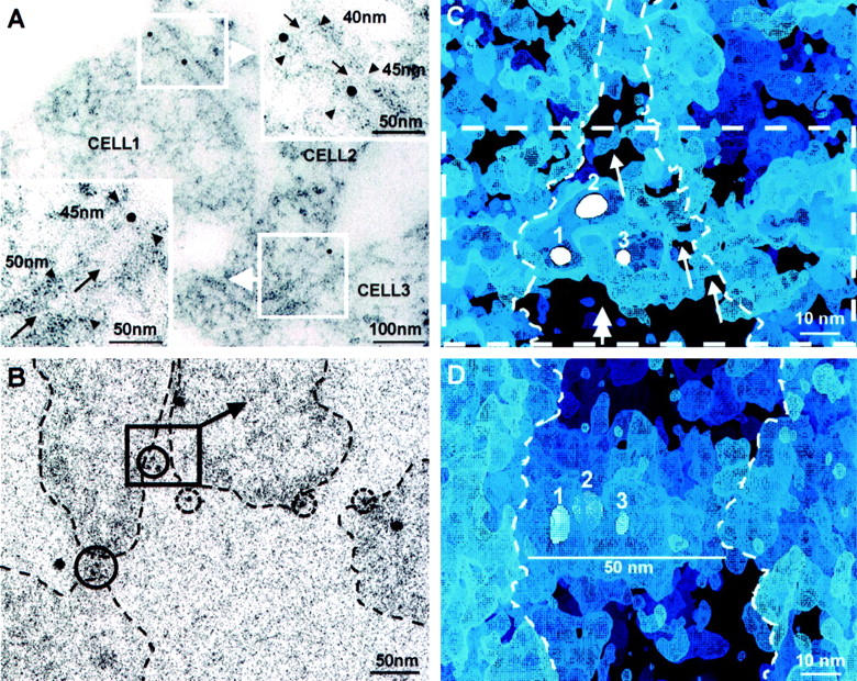
Nephrin-transfected HEK293 cells immunogold-labeled for extracellular nephrin. A: In horizontal EM thin section of a cell monolayer, nephrin immunolabel (10 nm gold) is seen at contacts between three cells (cells 1, 2, and 3). In blow-ups, cell membranes are ∼40 to 50 nm apart at sites of gold label, where also extracellular material is visible (arrows). B: Single low-dose image at 0° tilt from an electron tomography tilt series of sectioned cells as in A. Nephrin label (5-nm immunogold within solid circles) is seen concentrated at cell contact areas and solitarily (broken circles) on free cell surfaces. Cell boundaries marked by dashed lines. Volume chosen for three-dimensional visualization, shown in C, is marked by square. Large 10-nm unmarked gold particles are protein A-gold laid on grids for alignment purposes. C: A digital section of the electron tomography volume marked in B. Three immunogold particles (white spheres 1, 2, and 3, digital sections of gold particles located at different levels) mark sites of immunolabeled nephrin amino-ends in extracellular material between contacting cells. Thin ∼5-nm strands emanating from cell membrane are discernible in the labeled area (arrows). Strands border irregular pores in intercellular material. Dashed rectangle marks volume seen in D, viewed from indicated direction (double-headed arrow). Cell boundaries marked by dashed line as seen in stereo viewing. D: Slanted side-view of volume marked by dashed rectangle in C. A distinct layer of extracellular material bridges the cell boundaries (dashed lines). Immunogold particles (1 to 3) lie within the up to 50-nm wide extracellular layer. Cells cross-cut, growth substratum downward.