Figure 5.

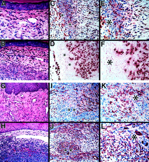
Comparison of lesions produced by 12E2 microinjection model and human Kaposi’s sarcoma. A and B: Conventional histology of 12E2 microinjection sites after 1 week, showing infiltration of superficial (A) and deeper (B) dermal layers by bland spindle cells associated with dilated blood vessels. C to F: Immunohistochemistry of adjacent sections of 12E2 microinjection site (1 week) stained for FXIIIa (C and E) and the endothelial marker, CD31 (D and F), documenting angiogenesis associated with infiltration of 12E2 cells. G and H: Conventional histology of characteristic human KS lesion. I to L: Immunohistochemistry of adjacent sections of typical human KS lesion stained for FXIIIa (I and K) and CD31 (J and L). Note histological and immunohistochemical similarities to the 12E2 microinjection sites (A to F). *Corresponding adjacent foci in C to F, I and J, K, and L FXIIIa/CD31 pairs. Original magnifications: ×400 (A, E to G, K, and L) and ×200 (B to D, H to J). H&E staining in A, B, G and H. NovaRed chromagen in C to F and I to L.
