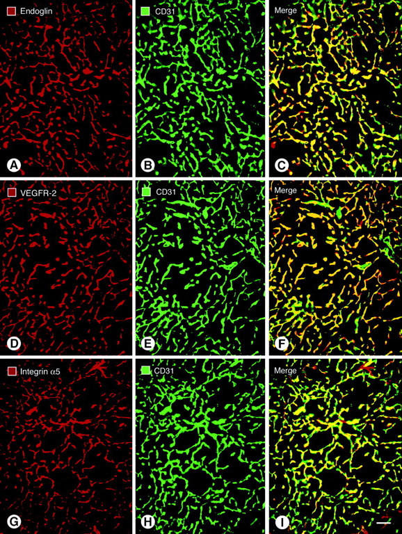
Figure 1.

Fluorescence micrographs showing blood vessels in RIP-Tag2 tumors double-stained for the endothelial cell markers endoglin (A); VEGFR-2 (D); or integrin alpha5 (G), shown in red; and CD31 immunoreactivity (B, E, H), shown in green. CD31 immunoreactivity primarily co-localizes with the other markers, but the overlap is not perfect (C, F, I). The most complete co-localization, indicated by yellow fluorescence in the merged image, is found with endoglin (C) and VEGFR-2 (F). Some vessels marked by CD31 have little or no integrin alpha5 immunoreactivity, as indicated by green fluorescence in the merged image (I). Few vessels lack CD31 immunoreactivity, as indicated by red fluorescence in merged images (C, F, I). Scale bar, 50 μm (A–I).
