
Figure 3.
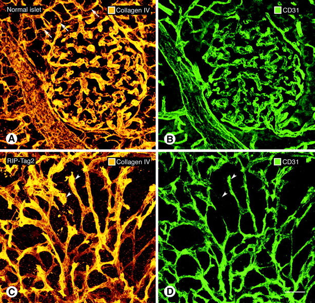
Confocal microscopic images of type IV collagen immunoreactivity of basement membrane (A, C) and CD31 immunoreactivity of endothelial cells (B, D) in normal pancreatic islet of wild-type mouse (A, B) and in RIP-Tag2 tumor (C, D). In both cases, all CD31-immunoreactive vessels co-localize with type IV collagen immunoreactivity. Tumor vessels are more irregular and tortuous but still have a uniform envelopment of type IV collagen immunoreactivity. Arrowheads mark tumor vessels where type IV collagen staining (C) is broader or more prominent than the corresponding CD31 immunoreactivity (D), suggestive of a loose association with endothelial cells. Arrows in A point to exocrine pancreatic acini outlined by type IV collagen immunoreactivity. Scale bar, 50 μm (A–D).