Figure 5.
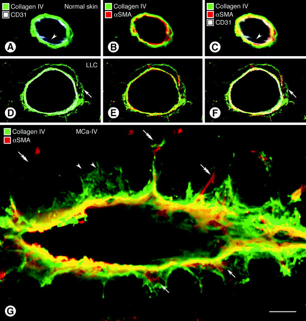
Confocal microscopic images comparing wall structure of 5-μm-thick optical sections of vessels in normal skin (A–C) and Lewis lung carcinoma (D–F, LLC) triple-stained for CD31, α-SMA, and type IV collagen immunoreactivities. Type IV collagen staining in the normal venule is closely apposed to CD31-immunoreactive endothelial cells (A) and α-SMA-immunoreactive pericytes (B). All three markers form a tight sandwich (C). Arrowheads mark endothelial cell nucleus. In the tumor vessel a layer of type IV collagen immunoreactivity extends well beyond the endothelial cells (D–F, arrows) and in some regions co-localizes with α-SMA-positive pericytes (E). A composite view (F) shows that the cell layers and basement membrane are separated by spaces (black). CD31 staining in some images (A, C, D, F) appears somewhat diffuse and not precisely limited to the plasma membrane for the reasons mentioned in the legend for Figure 4 ▶ . In G, multiple basement membrane abnormalities are shown in an oblique section through a vessel in MCa-IV breast carcinoma, double-stained for type IV collagen (green) and α-SMA (red) immunoreactivities, with regions of greatest overlap appearing yellow. Type IV collagen immunoreactivity spreads loosely from the vessel wall and penetrates the surrounding stroma (black). Extensions of type IV collagen immunoreactivity envelop α-SMA-immunoreactive pericytes in some regions (arrows) but not in others (arrowheads). Some α-SMA-immunoreactive cell processes extend away from the vessel wall (double arrows). Scale bar, 10 μm (A–G).

