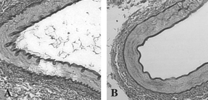
Figure 6.
Histopathological evaluation of renal artery from transgenic mice expressing human SSAO in smooth muscle cells. Cross-sections through renal artery were stained with the Weigert technique showing elastin in black. A: The renal artery displaying undulated internal and external elastic laminas from a non-transgenic littermate, with symmetrically arranged elastic fibers in tunica media. B: SMP8-SSAO transgenic mouse of line 12 displaying the same straight phenotype of the elastic laminas as seen in aorta. Both images are photographed at ×60 magnification.

