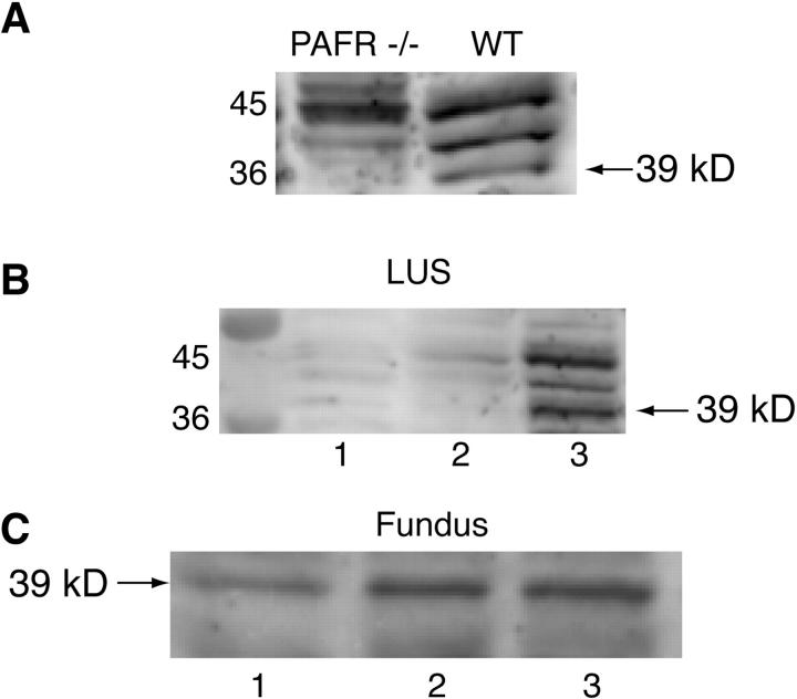
Figure 3.
Representative Western blots using the PAFR polyclonal antibody. A: PAFR antibody produces three bands in the 36 to 45 kd range. One of these bands at 39 kd is observed in pregnant uterine tissue from wild-type mice but not in pregnant uterine tissue from PAFR-/- mice. B: PAFR protein expression in the LUS in NP (lane 1), day 15 of gestation (lane 2) and day 19 of gestation (lane 3); a 39-kd band is visualized most prominently in day 19 uterine tissue. C: PAFR protein expression in fundal tissue: lane 1, NP; lane 2, day 15 of gestation; lane 3, day 19 of gestation.

