
Figure 4.
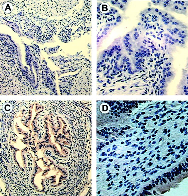
Immunohistochemistry using the PAFR antibody. A: Representative uterine section from PAFR-/- mice (×10). B: Uterine section from PAFR -/- mice at higher magnification (×20) demonstrating no antibody binding. C: Representative section from wild-type mice on day 15 of gestation at ×10 magnification. D: Representative section from CD-1 mice on day 15 of gestation at ×40. Moderate to intense staining is observed in the endometrial glands and smooth muscle, while lighter staining is seen in the smooth muscle only.