
Figure 1.
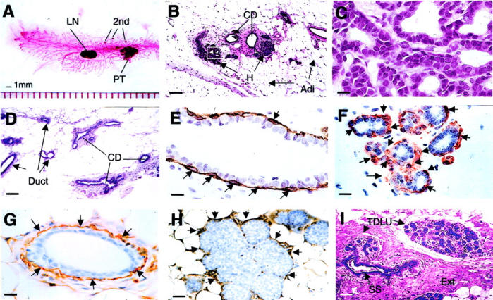
Early premalignant tumor in PyMT mice is comparable to human normal breast. A: Carmine red-stained whole-mount preparation of mammary gland from a PyMT mouse at 8 weeks of age. PT, primary tumor; second, foci growing on the distant ducts; LN, lymph note. B: H&E staining of a mammary gland prepared from a PyMT mouse at 4 weeks of age. CD, main milk-collecting ducts; H, the hyperplastic lesion; Adi, adipose cells. The inset in B is shown in C. D: H&E staining of a mammary gland section from a normal mouse at 4 weeks of age. E and F: IHC of SMA in a normal mouse mammary duct (E) and a hyperplastic lesion (F). Arrows point to the positively stained myoepithelial cells. G and H: IHC of laminin α1 in a normal mouse mammary duct (G) and a hyperplastic lesion (H). Arrows point to some of the positively stained areas. I: H&E staining of a normal human breast. TDLU, terminal duct lobular unit; SS, subsegmental duct; Ext, extracellular fibers. Note that SS leads to TDLU. The earliest change in the mouse lesion is an alteration in the mammary architecture that produces a lobular-alveolar structure that looks similar to the normal TDLU of the human. Scale bars: 100 μm (B, D, I); 25 μm (H); 10 μm (C, E, F, G). Original magnifications: ×100 (B, D, I); ×1000 (C, E, F, G); ×400 (H).