
Figure 7.
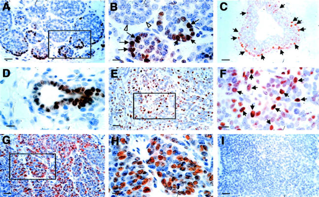
Increased expression of cyclin D1 during tumor progression to malignancy. A and B: Representative IHC of cyclin D1 in a PyMT-induced mouse mammary tumor at the adenoma/MIN stage (6 weeks of age). A: Positively stained cells are mainly found as a rim in the vicinity of the lesion. The inset in A is shown in B. Arrows point to cyclin D1-positive cells and open arrows point to adjacent acini that consist of mainly negatively stained cells. C and D: Cyclin D1 staining of an adjacent TEB and duct. Arrows point to positively stained cells. E: Cyclin D1 staining of a mammary tumor at the early carcinoma stage (9 weeks of age). The inset in E is shown in F. Arrows point to some of the positively stained cells. G and H: Staining of a primary tumor at the late carcinoma stage. The inset in G is shown in H. I: A mammary gland section stained with secondary antibody only as a negative control for the cyclin D1 IHC. Scale bars: 25 μm (A, C, E, G, I); 10 μm (B, D, F, H). Original magnifications: ×400 (A, C, E, G, I); ×1000 (B, D, F, H).