
Figure 3.
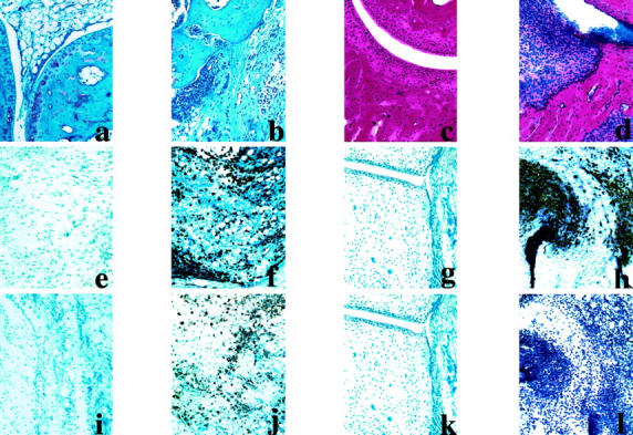
Histology of normal and arthritis joint sections and the infiltrated tissue surrounding the joints in CAIA. Top: Safranin staining of normal (a) and inflamed paw (b) of BALB/c mice on day 11 after antibody transfer (5 days after LPS injection) and hematoxylin staining of normal (c) and arthritis paw (d) of B10.RIII mice on day 17. Middle: Stained for RB6+ cells in normal and inflamed paws of BALB/c (e and f) and B10.RIII (g and h) mice, respectively. Bottom: Stained for Mac1+ cells in normal and inflamed paws of BALB/c (i and j) and B10.RIII (k and l) mice. e, f, h, i, j, and l: Surrounding tissues of the joints showing the nature of infiltrating immune cells in the inflamed paw. Results shown are representative of those obtained from three to four mice in each group. Original magnifications, ×20.