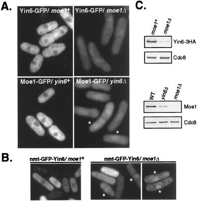
Figure 5.
Subcellular localizations and protein levels of Yin6 and Moe1. (A) The relevant genotypes of tested cells and the GFP-tagged proteins under investigation are indicated. Moe1-GFP concentrates more efficiently in the nucleus of yin6Δ cells; two such cells are marked by asterisks. The images were captured by the camera under identical conditions to reveal the drastically different intensities among these samples. As a control, we examined cells without a GFP tag and found autofluorescence signal that is primarily cytosolic and too weak to be photographed. The strains tested were ECP20 (yin6-gfp), ECP22 (yin6-gfp moe1Δ), ECP24 (moe1-gfp), and ECP25 (moe1-gfp yin6Δ). (B) yin6Δ or moe1Δ cells (YIN6K or MOE1U) transformed with pREP41GFPYIN6 were inoculated in thiamin-free MM medium and photographed. Asterisks mark those cells whose GFP-Yin6 is clearly concentrated in the nucleus. Cells were also transformed with a control vector expressing the GFP control, which diffuses throughout the cell (9). (C) The same amount of total proteins from each strain was analyzed by Western blotting to reveal the protein levels of Yin6-HA, Moe1, and Cdc8. The strains tested were, from the upper left to the lower right, ECP21 (yin6-HA moe1+), ECP22 (yin6-HA moe1Δ), SP870 (WT), YIN6K (yin6Δ), and MOE1L (moe1Δ).

