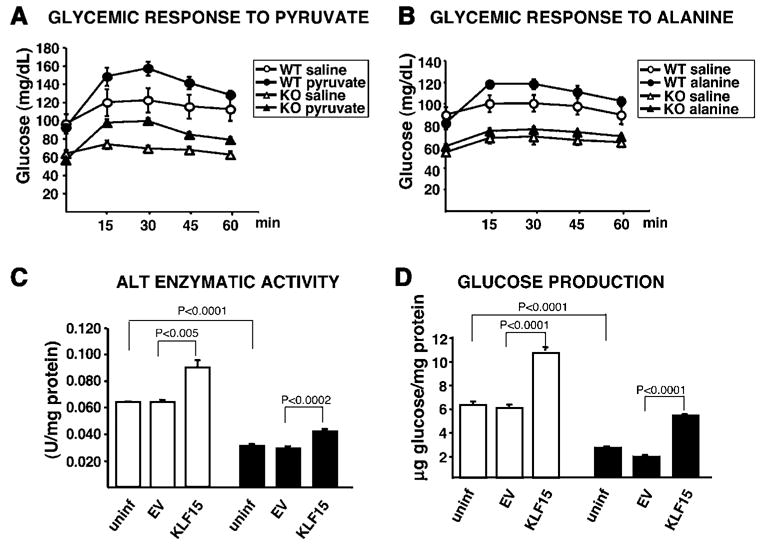
Figure 3. Pyruvate and Alanine Utilization In Vivo and ALT Activity and Glucose Production in Primary Hepatocytes.

(A and B) 18 hr-fasted WT and KLF15−/− (KO) mice each received an i.p. injection of 500 mg/kg sodium pyruvate dissolved in water or osmolarity-matched 104.5 mg/kg NaCl control solution (A) or 200 mg/kg L-alanine dissolved in 104.5 mg/kg NaCl or 104.5 mg/kg NaCl control solution (B). Tail-vein blood samples were assessed for glucose concentration immediately before injection (time 0) and at the indicated time points postinjection. n = 7 mice per condition. Statistical comparisons were made using MANOVA (p ≤ 0.05 was considered significant). p = 0.0318 WT saline versus WT pyruvate; p = 0.0034 KO saline versus KO pyruvate; p = 0.02647 WT saline versus WT alanine; p = 0.7843 KO saline versus KO alanine.
(C and D) WT (white bars) and KLF15−/− primary hepatocytes (black bars) were either uninfected or adenovirally infected at 30 moi with EV or KLF15 constructs (~10-fold overexpression of KLF15) and incubated at 37°C in glucose production buffer (see Experimental Procedures). The buffer was removed after 3 hr and assayed for glucose content, and the cell lysate was assayed for ALT activity. n = 4 replicates per group for each assay. Statistical comparisons were made using Student’s t test for unpaired samples.
