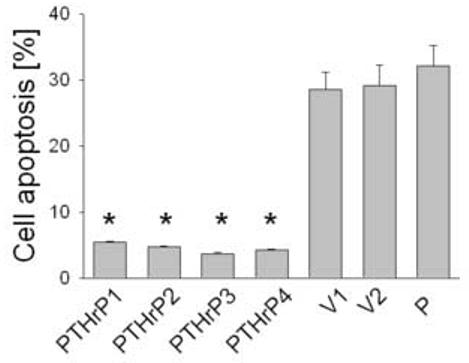
Figure 4.

Apoptosis of LoVo cells after modulating PTHrP expression. Apoptosis was measured after staining with Annexin V. Cells were plated in medium containing 10% FBS, then transferred to medium containing 0.1% FBS after 48 h. Apoptosis was measured after 16 h as described in Materials and Methods. Data for each of four independent PTHrP-overexpressing clones (PTHrP1 to PTHrP4), two independent empty vector-transfected clones (V1 and V2; controls), and parental cells (P) are presented. The PTHrP-overexpressing clones secrete between 55-fold and 75-fold more PTHrP than the empty vector-transfected cells. Each bar is the mean ± SEM of three independent experiments. * = Significantly different from the control value (P < 0.001).
