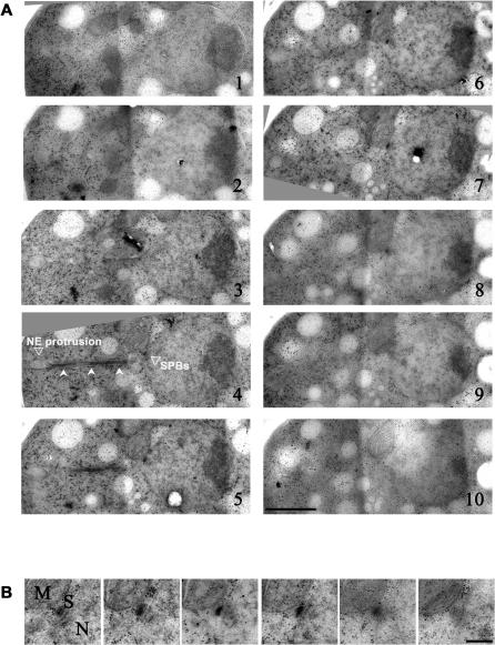
Figure 3. The SPBs Are Duplicated but Not Separated in Cells Overexpressing Mia1p.
(A) Electron microscopy images of serial sections of a Mia1p-overexpressing cell showing the abnormal panhandle-shaped protrusion of the NE. Note that microtubules extend throughout the protrusion (indicated by indented arrowheads, panel 4). The SPB pair is located at the base of protrusion. The NE protrusion and position of the SPBs are indicated on panel 4. Scale bar represents 1 μm.
(B) Higher magnification image of the duplicated SPB pair. Mitochondrion is labeled as M, nucleus as N. Scale bar represents 0.2 μm.

