
Fig. 3.
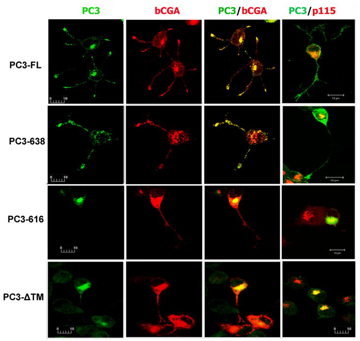
Subcellular distribution of PC3 in transfected N2A cells by fluorescence immunocytochemistry and confocal microscopy. N2A cells were co-transfected with four PC3 constructs and an expression construct encoding bovine chromogranin A (bCGA). The green signal indicates immunoreactive PC3 (1st column) and the red signal indicates bCGA, the secretory granule marker, in transfected cells (2nd column). The merged confocal images show the co-localization of PC3 and bCGA (yellow, in 3rd column) in the cell processes of full length PC3 (PC3-FL) and carboxyl terminus truncated PC-638 transfected cells. The 4th column shows co-localization of PC3 (green) and p115 (red) in transfected cells. The PC3 staining in PC3-616 or PC3-ΔTM transfected cells was primarily present in the cell body and colocalized with p115, a Golgi marker. The bar represents 10 μm.