Figure 1.
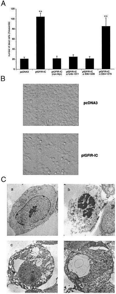
The IGFIR-IC induces cell death. (A) Comparison between the cell death induced by pIGFIR-IC and nonmyristylated form of the IC domain and deletion mutants. Asterisks indicate a highly significant difference from the control as determined by one-way ANOVA (F = 63.3; P < 0.0001); P < 0.0001 for pIGFIR-IC and pIGFIR-ICΔ1264–1276, Bonferroni/Dunn posthoc test. Error bars represent the SEM from three independent experiments. (B) Light microscopy, using Hoffman optics, demonstrating morphological changes induced by IGFIR-IC. IGFIR-IC expression caused a rounding of the cells and intracellular vacuoles. (C) Ultrastructural characteristics of IGFIR-induced cell death. 293T cells transfected with control empty vector pcDNA3 (a), pC9 (b), or pIGFIR-IC (c and d). Although caspase-9-transfected cells displayed characteristic features of apoptosis, including chromatin condensation, IGFIR-IC-transfected cells did not. In contrast, extensive cytoplasmic vacuolization was observed in the absence of nuclear fragmentation, cellular blebbing, or apoptotic body formation. Note that the cell shown in b displays both the chromatin condensation characteristic of apoptosis and membrane disruption characteristic of necrosis, suggesting secondary necrosis after apoptosis. (Original magnification: ×6,000.)

