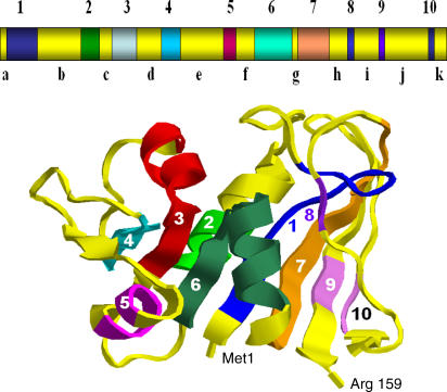
Figure 2. A Drawing of the Tertiary Structure of E. coli DHFR Taken from the Protein Data Bank and the Locations of the Proposed Folding Elements [5].
Four α-helices and eight β-strands are shown as ribbons. Proposed folding elements are light blue (1: Ser3~Ile14), green (2: Trp30~Leu36), red (3: Val40~Ser49), cyan (4: Arg57~Ser63), magenta (5: Glu80~Ala83), greenish-blue (6: Ile91~Leu104), orange (7: Ala107~Glu118), purple (8: His124~Phe125), and light purple (9: Val136~Ser138; and 10: Glu154~Ile155), respectively. The regions outside the folding elements form amino acid segments a–k (in yellow). The positions of the N- and C-termini are also indicated. Also see Table 1.

