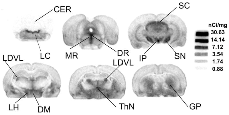
Figure 3.

Ex vivo autoradiographic localization of [125I]7 binding sites in rats 4 hours post-injection in a normal rat – High levels of radioactivity were observed in areas containing high densities of serotonin transporter sites. Rat brain sections showed intense labeling in several regions [46], i.e. lateral hypothalamic area (LH), dorsomedial hypothalamic nucleus (DM), interpeduncular nucleus (IP), thalamus nuclei (ThN), dorsal raphe (DR), medial raphe (MR), superior colliculus (SC), locus coeruleus (LC), sustantia nigra (SN), globus pallidus (GP), areas known to have high densities of SERT sites [47]. Lower, but detectable, labeling was also found in the frontal cortex, caudate putamen, ventral pallidum and hippocampus, areas containing a significantly lower number of SERT sites. The coronal sections correspond to the stereotaxic atlas [46].
