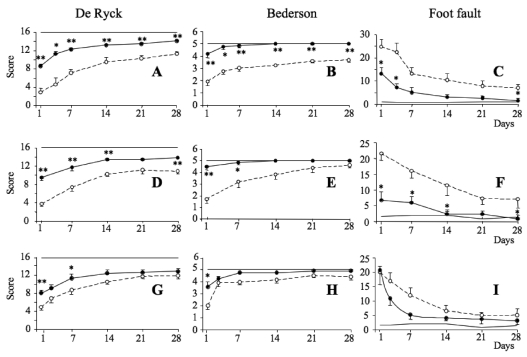
Figure 4.
Effect of reparixin treatment under different schedules on neurological deficits after transient MCAO. Transient (1.5 h) MCAO was performed. Reparixin was administered as follows: Panels A-C: 15 mg/kg, i.v. 2 h after reperfusion (i.e. 3.5 h after MCAO) followed by two 15 mg/kg s.c. doses at 2 h intervals, then 3 times/day on days 2 and 3. Panels D-F: 15 mg/kg, i.v. 2 h after reperfusion (i.e. 3.5 h after MCAO) followed by s.c. infusion for 48 h at a rate of 10mg/h/kg. Panels G-I: 15 mg/kg, i.v. 4.5 h after reperfusion (6 h after MCAO) followed by s.c. infusion for 48 h at a rate of 10mg/h/kg. Neurological deficits were evaluated by De Ryck’s (left panels), Bederson’s (middle panels) and foot-fault (right panels) tests starting from 24 h after MCAO. Open circles, vehicle; closed circles, reparixin. For comparison, the scores of non-ischemic, sham-operated rats, i.e. “normal” values, are also shown as a thin line (normal values were: 16 for the De Ryck’s test, 5 for the Bederson’s test and 1–4 for the foot-fault test. Data are mean ± S.E. of 6–7. Statistical differences (Mann-Whitney): *P < 0.05 and **P < 0.01 vs. saline.

