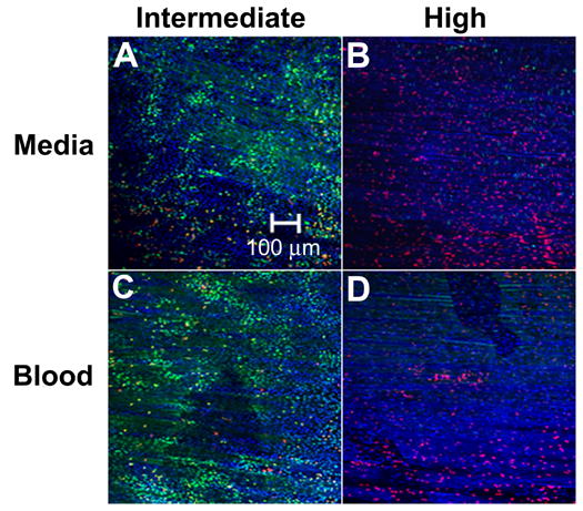
Figure 6.

Confocal microscopy images of endothelium displaying bioeffects mediated by ultrasound exposure to intact arteries at near physiologic conditions. Ultrasound was applied at (A, C) intermediate and (B, D) high energy while the artery was filled with (A, B) DMEM or (C, D) blood.
