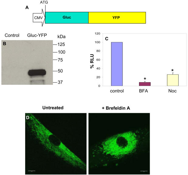
Figure 4. Gluc-YFP fusion to visualize secretory pathway in real-time.
(A) Schematic representation of the Gluc-YFP fusion cloned in the lentivirus vector. (B) Uninfected 293T cells or cells infected with a lentivirus vector expressing Gluc-YFP fusion were lysed and analyzed by western blotting with anti-Gluc antibody. (C) Cells expressing Gluc-YFP fusion were treated with BFA or nocodazole and their conditioned medium were assayed for Gluc activity 24 hrs later. *p≤0.01 as predicted by student T-test. (D) Fluorescence microscopy of a single live cell expressing Gluc-YFP and either untreated or treated with BFA showing that this fusion is trapped in the ER upon BFA treatment. Scale bar, 10 µm.

