
Figure 4.
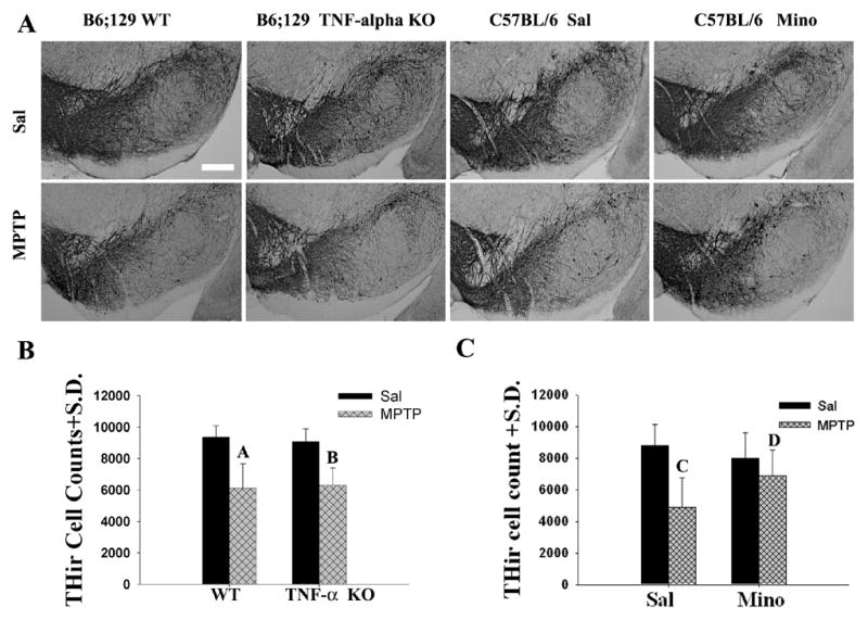
The loss of dopamine neurons was attenuated by minocycline treatment, not by TNF-α KO following MPTP treatment. (A) Photomicrographs of representative sections of THir staining in the SNs of the mice used to generate the bar graph of (B) and (C). SN of (a) WT/Sal, (b) TNF-α KO/Sal, (c) WT/MPTP, (d) TNF-α KO/MPTP, (e) Sal/Sal, (f) Mino/Sal, (g) MPTP/Sal, and (h) MPTP/Mino (Magnification bar=0.25mm). (B) Quantification of THir cells in the WT and TNF-α KO mice following MPTP treatment. MPTP similarly reduced the numbers of THir cells in the SN of both WT and TNF-α KO mice. The number of THir cells was not significantly different in WT/MPTP and TNF-α KO/MPTP mice [(p=0.774); Marked A and B, separately]. (C) Quantification of THir cells in C57BL/6 mice with or without minocycline treatment following MPTP administration. The number of THir cells was significantly different (p=0.044) between Sal/MPTP (Marker C) and Mino/MPTP mice (Marker D).