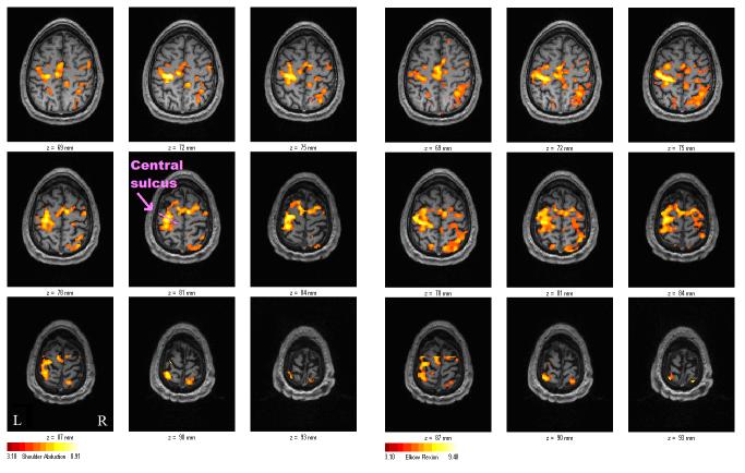
Figure 6.
The subject's activation maps for right-handed shoulder abduction task (left) and right-handed elbow flexion task (right). The primary motor cortices, supplementary motor area, premotor areas, and primary sensory area were activated under both conditions. Comparison between the two tasks in the single subject yielded no statistically different activation sites (p > 0.05).

