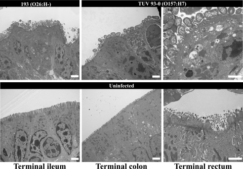
FIG. 3.
Representative transmission electron microscopy micrographs showing EHEC O26:H− (193 Nalr) and O157:H7 (TUV 93-0) inducing typical A/E lesions in the terminal ileum, colon, and rectum. A/E lesions were characterized by intimately adherent bacteria, effacement of the brush border, and F-actin accumulation underneath adherent bacteria. No A/E lesions were observed on uninfected sections. Bar = 2 μm.

