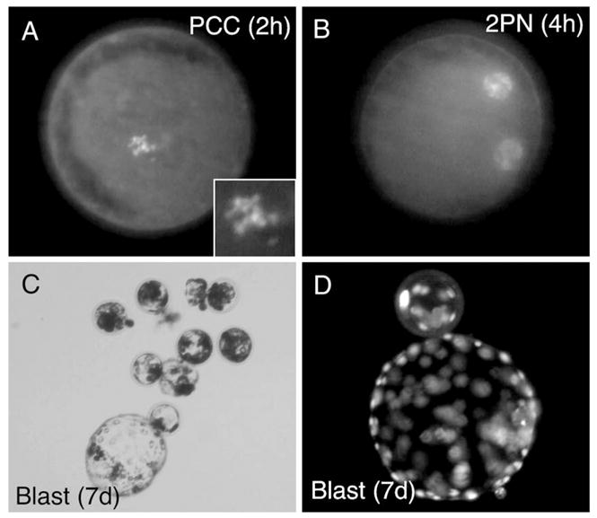
Fig. 3.

In vitro development of fibroblast NT embryos following cell fusion and activation. (A) DAPI staining of PCC formation at 2 h post-SCNT. (B) DAPI staining of pronuclei formation at 4 h post-SCNT. (C) Bright field photomicrograph of 7-day blastocysts formed following SCNT. (D) DAPI stained 7-day blastocysts formed following SCNT.
