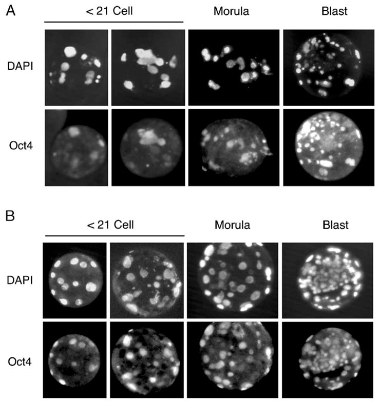
Fig. 4.

Oct4 staining of parthenogenetically activated oocytes. Oocytes were collected (A) following eCG treatment of donor Jills or (B) following natural mating with vasectomized males. Oocytes were activated as described in Materials and methods and stained with anti-Oct4 antibodies and DAPI at various stages as indicated. A total of four Jills were used to generate at least 6 embryos in each developmental group. Oct4 expression was seen in 47 ± 14% of nuclei from ≤21 cells embryos of the hormonally treated group and 83 ± 5% of nuclei from ≤21 cells embryos in the untreated group.
