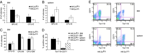
Fig. 3.
Erythroid progenitor cells are decreased in Hif-2αΔ/Δ bone marrow but increased in the spleen. (A) Hif-2αΔ/Δ bone marrow progenitor cells form fewer erythroid colonies in methylcellulose than Hif-2αfl/Δ cells do (n = 4). (B) In contrast, Hif-2αΔ/Δ splenic progenitor cells have a higher potential to form erythroid colonies than cells from Hif-2αfl/Δ spleens (n = 4). (C) There is little difference in the number of nonerythroid colonies formed by Hif-2αfl/Δ and Hif-2αΔ/Δ bone marrow cells (n = 4). (D) The percentage of Ter119high/CD71high erythroid progenitor cells is reduced in Hif-2αΔ/Δ bone marrow, but increased in Hif-2αΔ/Δ spleen (n = 3). (E) Representative FACS blots for Hif-2αfl/Δ and Hif-2αΔ/Δ bone marrow and spleen stained for Ter119 and CD71.

