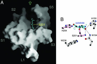
Fig. 3.
A hypothetical model of bound substrate peptide. (A) The peptide (cyan) is positioned in the open trough at the top of the membrane protease, with its carboxyl terminus pointing toward the gap between transmembrane helices S2 and S5. (B) A detailed view of the hypothetical substrate (cyan) bound to the GlpG active site. This picture corresponds to a view from the back of GlpG as indicated by the green arrow in A. The carbonyl carbon atom of the scissile bond is positioned just above the hydroxyl group of Ser-201, and the carbonyl oxygen atom occupies the proposed oxyanion-binding site defined by the main chain of Ser-201 and the side chains of His-150 and Asn-154.

