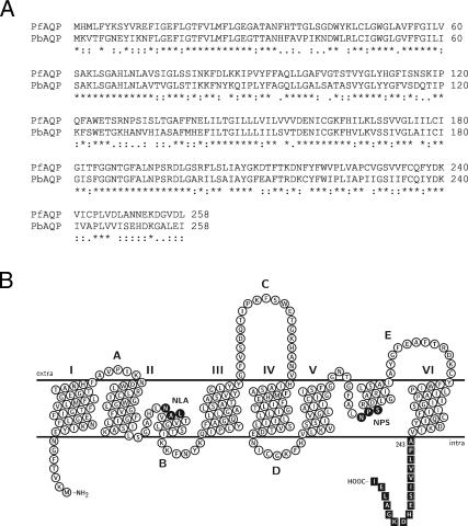
Fig. 1.
Comparative alignment and predicted topology of PbAQP. (A) Alignment of PbAQP with PfAQP was performed with ClustalX 1.81. Asterisks, colons, and periods indicate identical, highly, and moderately conserved amino acid residues, respectively. (B) Predicted membrane topology of PbAQP. The putative transmembrane domains were assigned by hydrophobicity analysis and by manually threading the sequence of PbAQP through the x-ray crystal structure of E. coli GlpF (Protein Data Bank ID code 1FX8). The topology map was drawn with TeXtopo. NLA and NPS motifs are indicated. The peptide used for antibody production is indicated by boxes.

