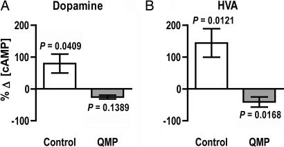
Fig. 5.
Responses of isolated mushroom body calyces to dopamine monitored by using measurements of intracellular cAMP. Calyces from QMP-exposed bees (gray bars) and from control bees (white bars) were exposed to either 10 μM dopamine (A) or 10 μM HVA (B). Note that dopamine-evoked responses are strikingly different in QMP-treated bees versus controls and that the effects of dopamine are mimicked by HVA. Data are expressed as mean levels ± SEM with a sample size of six for each group. P values refer to within-group differences between cAMP levels detected in dopamine-treated or HVA-treated tissues and those detected in tissues that were not exposed to dopamine or HVA.

