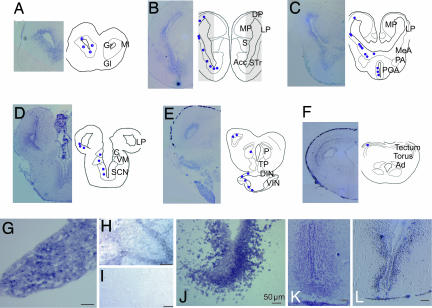
Fig. 3.
LHR mRNA expression in the Xenopus brain detected by in situ hybridization. Photomicrographs of transverse sections through Xenopus brain from anterior (A) through posterior (F). (A–F) Left columns of the sections show LHR mRNA expression as a purple precipitate, whereas Right columns show schematic drawings of brain with purple dots illustrating the positive hybridization signal. High magnification photomicrographs of the striatum (J), POA (K), and ventral hypothalamus (L) show LHR mRNA expression. Strong LHR mRNA hybridization signal was shown in pituitary (G), and Leydig cells in testis (H). RNase pretreatment eliminates hybridization (I). (Scale bar: 50 μm.) Nomenclature is as in refs. 57 and 58. For abbreviations, see abbreviations list.

