
Figure 5. Effect of IFN-γ on division of donor CD8 T cells.
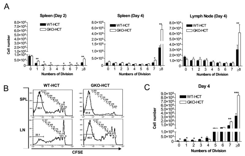
A–B. B6D2F1 mice received BMC and CFSE-labeled CD4-depleted spleen cells from WT (closed bars) or GKO (open bars) B6 donors (n=3 per group). Recipient spleen and lymph node cells were prepared at the indicated time points and analyzed for division of donor CD8 T cells. A. Numbers of donor CD8 T cells with each division in the spleen at days 2 and 4, and in the lymph nodes at day 4 post-HCT. Axillary, brachial and inguinal lymph nodes were harvested at indicated time point, and pooled lymph node cells were analyzed. B. Representative histograms showing CFSE levels in gated donor CD8 T cells at day 4. Numbers indicate the percentages of cells with each number of cell divisions. C. B6D2F1 mice received BMC and CFSE-labeled spleen cells from WT (closed bars) or GKO (open bars) 2C donors (n=3 per group). Data shown are the percentages of donor 2C TCR+ cells with different cell divisions (mean±SDs) in the recipient spleen at day 4 post-HCT. *, p<0.05; **, p<0.01, ***, p<0.001.