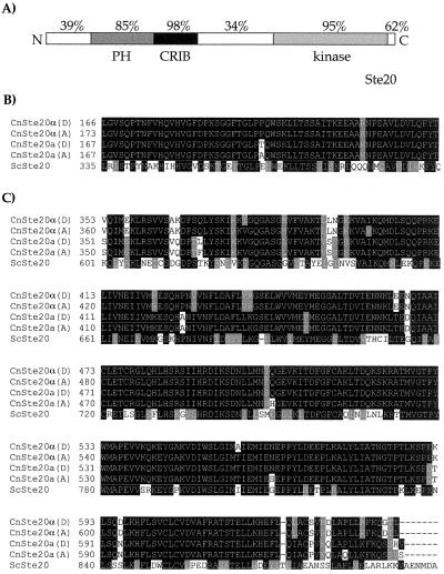
Figure 1.
Protein sequence alignment of Ste20 kinases of C. neoformans. The amino acid sequences of four Ste20 kinases from C. neoformans and their S. cerevisiae homolog were aligned by using the clustalw algorithm. (A) Schematic drawing of the Ste20 protein of C. neoformans depicting a putative (pleckstrin homology) domain, the Cdc42-binding domain (CRIB), and the carboxyl-terminal kinase domain. Percent values indicate the similarity between the serotype D Ste20a and Ste20α proteins in the conserved domains and the linker regions. Similar results were obtained comparing the serotype A-specific proteins. (B) Alignment of the CRIB domains. (C) Alignment of the carboxyl-terminal kinase domains. Amino acids present in at least two of five proteins were shaded. Black shading indicates identical amino acids, and gray shading indicates conserved residues. (See also Figs. 7–11, which are published as supplementary material at http://www.duke.edu/∼lengeler/PNAS.html.)

